Section the paraffin embedded tissue block at 5 8 µm thickness on a microtome and float in a 40 c water bath containing distilled water.
Immunofluorescence protocol for paraffin embedded sections.
Before moving to alcohol grades step make sure to completely deparaffinize the sections.
Mount the sections onto either probeon or probeon plus or gelatin coated histological slides.
If the sections still have traces of wax an additional immersion of 5 minutes in xylene may be employed.
Float the sections in a 56 c water bath.
Cut 5 15 µm thick tissue sections using a cryostat.
If it is too warm it will stick to the knife.
Cut 5 15 µm thick tissue sections using a rotary microtome.
The suggested cryostat temperature is between 15 and 23 c.
Immunohistochemistry protocol for paraffin embedded tissue sections advertisement doi.
The paraffin tissue block can be stored at room temperature for years.
Immunofluorescence general protocol important.
Studies performed as early as the 1970s have shown that if p is a valuable salvage technique in renal pathology when frozen tissue is inadequate e g lacks glomeruli or not available 8 10 14 18 20 21 overall diagnostic results by if p can be obtained in 80 of cases 14 18 20 21 but the diagnostic yield varies depending on 3.
Do not use this pretreatment with frozen sections or cultured cells that are not paraffin embedded.
Antigenic determinants masked by formalin fixation and paraffin embedding often may be exposed by epitope umasking enzymatic digestion or saponin etc.
Embed the tissue in paraffin at 58 c.
Tissues can be embedded into paraffin using specialized automated tissue processing systems.
Slides are pre coated with gelatin to enhance adhesion of the.
April 10 2014 katie crosby 1 jessica simendinger 1 christopher grange 1 michelle ferrante 1 trisha bernier 1 claire standen 1.
Please refer to the applications section on the front page of product datasheet or product webpage to determine if this product is validated and approved for use on cultured cell lines if ic paraffin embedded samples if p or frozen tissue sections if f.
Transfer the sections onto glass slides suitable for immunohistochemistry e g.
Embed the tissue in a paraffin block.
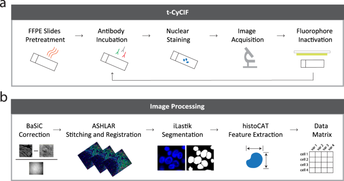
Download immunofluorescent staining of paraffin embedded tissue protocol as a pdf deparaffinization and rehydration tip.
A fluorescent dye is a fluorophore that is a molecule that absorbs light energy at a specific wavelength by a process called excitation and then immediately releases the energy at a different wavelength known as emission.
Ihc protocol video for paraffin embedded tissue sections from cell signaling technology cst cst protocols.
Thaw mount the sections onto gelatin coated histological slides.
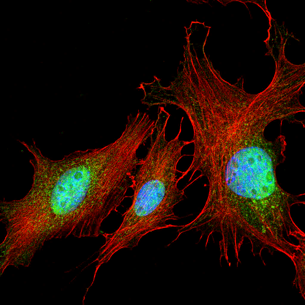
Immunofluorescence microscopy is a method by which a protein can be visualized inside cells using fluorescent dyes.


